OB ultrasound
33 studies11 challenges
Where is the pregnancy, how old is it, is it well: the questions each image answers, in the order the clinic asks them. Every image opens with how to read it; switch to Practice when you are ready to be tested.
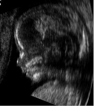 Mid-sagittal profile · 14 weeks
OB ultrasound · Anatomy scan
Fetal profile at fourteen weeks
The plane, and which way is up
Transabdominal at 18 to 22 weeks, in standard planes: the transthalamic and transcerebellar planes of the head, the four-chamber heart, the abdominal circumference plane with the stomach and umbilical vein, the femur, the sagittal spine and the face in profile and coronal view.
Look for, in order
- Head: skull shape and ossification, the midline, the cavum septi pellucidi, the ventricles (atrium under 10 mm), the cerebellum and cisterna magna (under 10 mm), the nuchal fold.
- Face: the profile with the nasal bone, the lips in coronal view, the orbits.
- Spine: intact skin line and vertebral arches in sagittal, transverse and coronal planes.
- Heart: situs, four chambers of equal size, the crux, the outflow tracts crossing.
- Abdomen and limbs: stomach on the left, kidneys and bladder, a three-vessel cord, the long bones and the hands and feet.
- Then the placenta, the cervix, the fluid and the biometry.
What normal looks like
Each plane has a checklist of structures; a normal scan is a scan where every one was seen. Biometry within range for the dates; fluid normal; placenta clear of the os.
Trap. A structure not seen is not a structure that is normal. Record what could not be seen and bring her back.
Describe it in this order
- The plane and the approach (transabdominal or transvaginal)
- Location of the pregnancy: in the uterus or not
- Number: singleton or multiple, and chorionicity
- Viability: cardiac activity
- Dating or growth: the measurement that fits the stage
- Fetal anatomy, placenta, cord, fluid, cervix, as the stage allows
- Adnexa and free fluid
- Then, and only then, the impression
Findings
- A normal profile with forehead, nasal bone, lips and chin in line
- No micrognathia, no frontal bossing
What it means for the patient
The profile is where the nasal bone, the facial angle, the lips and the chin are assessed. A receding chin (micrognathia) or a flat profile prompts a closer look at the face and a search for associated anomalies.
Framework: ISUOG, Practice guidelines: first-trimester and mid-trimester fetal ultrasound (2022).
OBGynSpace Tutor
It coaches the description; it does not read the image for you and will not name the diagnosis before you commit.